هل يمكن الشفاء من نزيف المخ

Can Brain Hemorrhage Be Cured?

Can brain hemorrhage be cured?
Recovery from a brain hemorrhage is possible in many cases, especially with rapid medical intervention and a carefully planned treatment approach. The extent of recovery depends on several key factors that directly affect healing outcomes, such as the size of the hemorrhage, its location in the brain, the patient’s age, and overall health condition.

Outcomes vary from one patient to another. Some cases show gradual improvement with near-complete restoration of neurological functions, while others may experience permanent complications requiring long-term rehabilitation.

In this article, we will review the factors affecting recovery, the main treatment options available, and the steps that can be taken to support healing and safely improve the chances of regaining health.

Brain Hemorrhage:

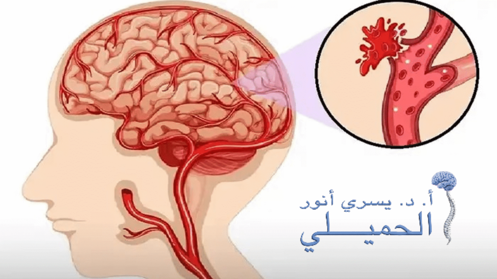
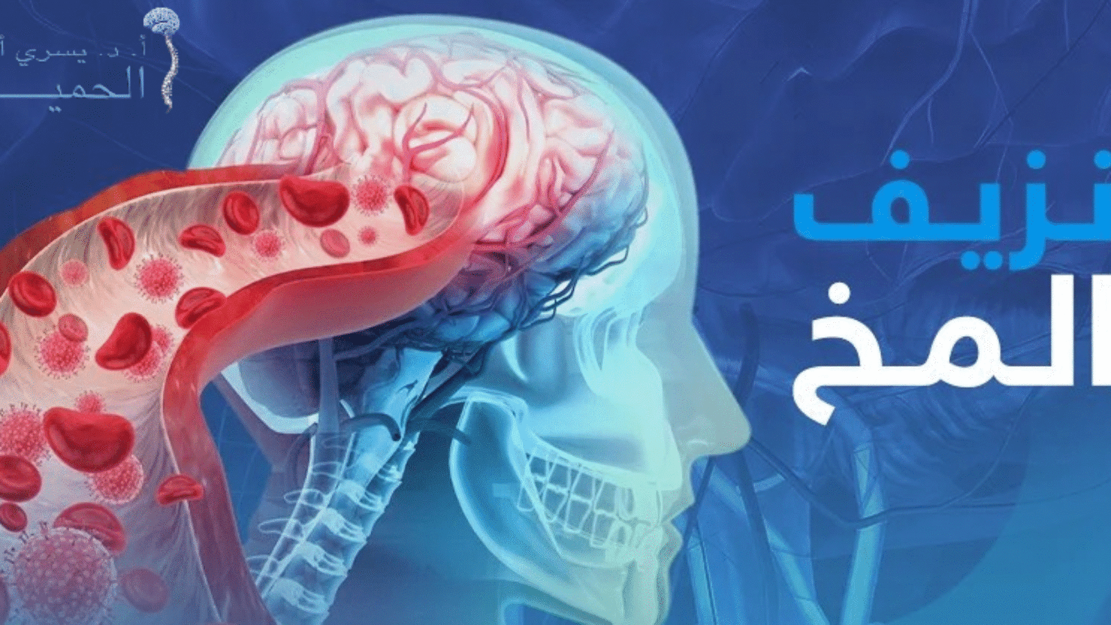
A brain hemorrhage is a form of stroke that occurs when blood vessels in the brain rupture, often due to a sudden rise in blood pressure. This leads to blood leaking into brain tissue, reducing blood flow and nutrient supply to affected areas and limiting oxygen delivery essential for normal brain cell function. As a result, brain cells may gradually die, and in some cases, the condition can progress to partial or complete paralysis if not treated urgently or if complications are not controlled in time.

Causes:

Several factors can lead to a brain hemorrhage, and identifying the exact cause often guides treatment decisions. The most common causes include:

  • Chronic high blood pressure: Continuous pressure weakens blood vessel walls and increases the risk of rupture.

  • Head injuries and trauma: Falls or traffic accidents can cause intracranial bleeding, particularly in children and young adults.

  • Aneurysms: Weakness in brain artery walls may lead to rupture and internal bleeding.

  • Congenital vascular malformations: Such as arteriovenous malformations (AVMs), where blood vessels are more prone to bleeding from birth.

  • Brain tumors: Some tumors, especially malignant ones, may cause bleeding due to pressure on blood vessels or vessel rupture.

  • Blood and clotting disorders: Low platelet counts or clotting disorders increase bleeding risk.

  • Chronic diseases: Conditions such as diabetes, heart disease, and atherosclerosis affect blood vessel health.

  • Lifestyle factors: Smoking, alcohol consumption, and drug abuse weaken blood vessels.

  • Pregnancy and childbirth complications: Conditions such as preeclampsia or the effects of certain contraceptives may increase hemorrhage risk.

  • Blocked brain arteries or clots: Arterial blockage can raise pressure and cause blood leakage.

Can Brain Hemorrhage Be Cured?

The possibility of recovery depends on multiple factors related to the patient’s condition and the severity of the hemorrhage. In some cases, full recovery is achievable, while in others, long-term complications affecting brain function may remain. The earlier the medical intervention and the more limited the brain tissue damage, the higher the chances of recovery.

Factors Affecting Recovery:

  • Location of the hemorrhage: Bleeding near vital brain areas makes recovery more challenging.

  • Size of the hemorrhage: Larger hemorrhages increase the likelihood of permanent brain damage.

  • Speed of medical intervention: Early diagnosis and prompt treatment significantly improve recovery chances.

  • Overall health condition: Patients with chronic illnesses may face greater recovery challenges.

Treatment Options:

Types of Treatment for Brain Hemorrhage

Treatment depends on the size and location of the hemorrhage and the patient’s condition:

1. Medical Treatment:

  • Medications to lower blood pressure.

  • Drugs to control brain swelling and prevent complications.

2. Surgical Treatment:

  • Removal of accumulated blood to relieve pressure on the brain.

  • Repair of damaged blood vessels.

  • Use of endoscopic techniques to minimize surgical intervention.

3. Rehabilitation After Hemorrhage:

  • Physical therapy to improve movement and balance.

  • Occupational therapy to support daily activities.

  • Psychological support to manage stress and anxiety.

4. Supportive Care:

  • Close medical follow-up to prevent complications and ensure treatment effectiveness.

Is Full Recovery Possible After a Brain Hemorrhage?

This depends on each patient’s individual condition. Some patients may return to their normal lives after full recovery, while others may experience long-term effects such as difficulty with movement, speech, or concentration. In such cases, physical and psychological rehabilitation becomes essential to improve daily functioning and adapt to post-hemorrhage challenges.

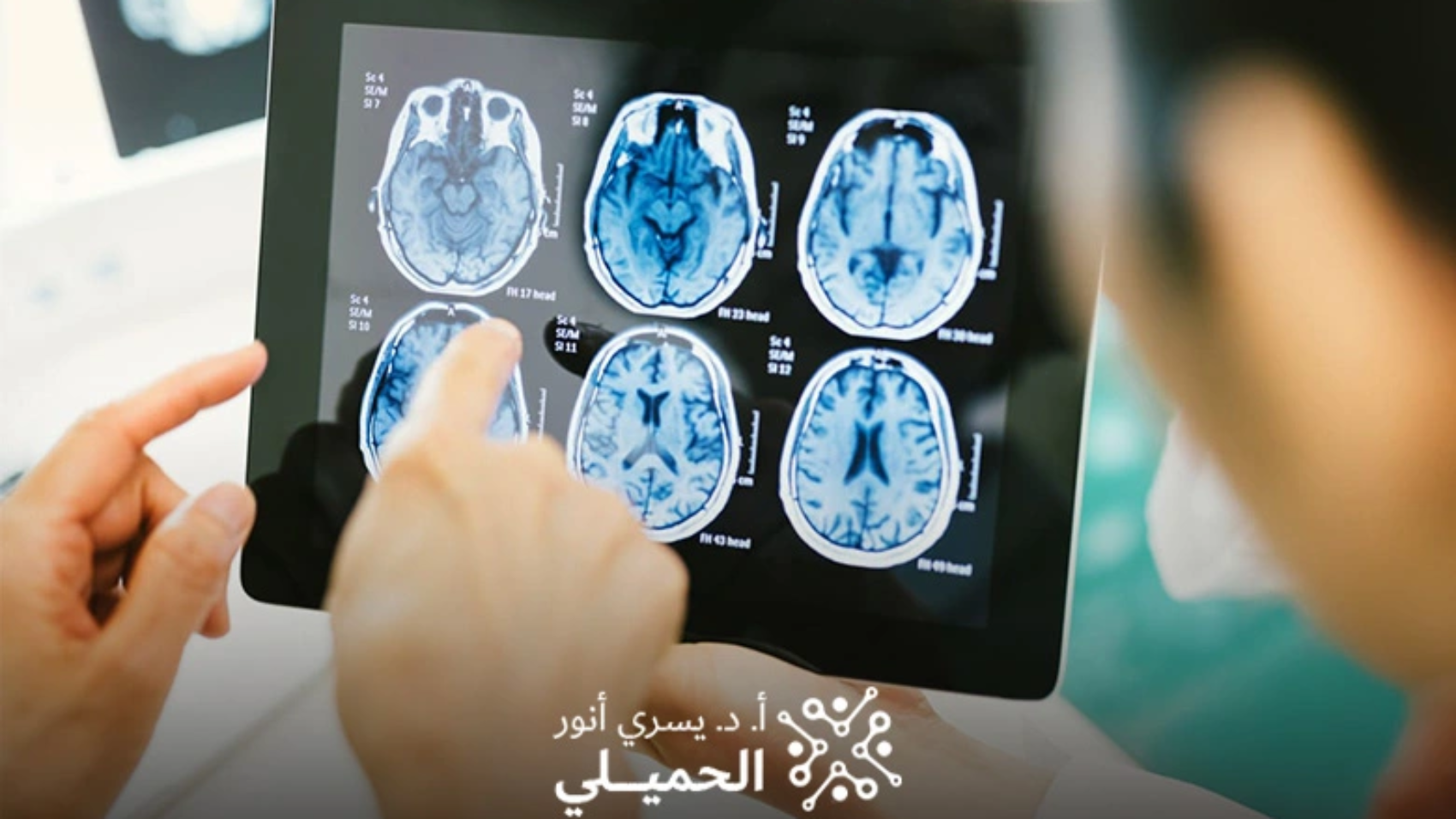
How Is Brain Hemorrhage Diagnosed?

Severe brain injuries are medical emergencies that require rapid assessment and treatment. The physician examines the patient and asks key questions, such as whether a head injury occurred, the specific symptoms experienced, whether the patient lost consciousness, and for how long. Based on this information, the doctor determines the location and size of the hemorrhage and conducts diagnostic tests, including:

  • CT scan of the brain

  • MRI of the brain

How Can Brain Hemorrhage Be Prevented?

The risk of brain hemorrhage can be reduced by following simple and effective measures, including:

  • Monitoring blood pressure: Keeping blood pressure within normal ranges reduces vessel rupture risk.

  • Preventing injuries: Using safety equipment during sports or driving to avoid head trauma.

  • Quitting smoking: Smoking increases blood vessel fragility.

  • Following a healthy diet: Balanced nutrition supports cardiovascular and vascular health.

When Should You Consult a Brain Hemorrhage Specialist?

If you need specialized medical advice regarding brain hemorrhage and its aftereffects, Dr. Yousri Al-Hamili is considered one of the leading specialists in neurosurgery, offering expertise in managing brain hemorrhage cases and guidance on neurological care after bleeding.

You should seek immediate medical attention if any of the following symptoms appear:

  • Sudden, severe headache unlike usual headaches

  • Weakness or numbness in one side of the body

  • Difficulty speaking or seeing

  • Loss of consciousness or decreased alertness

Early diagnosis and rapid intervention play a crucial role in reducing the risk of serious complications and improving recovery outcomes.

Conclusion

Recovery from a brain hemorrhage depends on the speed of medical intervention, the type of hemorrhage, and the patient’s overall health. Some patients achieve full recovery, while others may require long-term follow-up to manage lasting effects. Adherence to treatment plans and regular follow-up with specialized physicians are key to achieving the best possible outcomes.

Frequently Asked Questions (FAQ)

1. Can brain hemorrhage be cured?
Yes, recovery from a brain hemorrhage is possible in many cases, especially with early medical intervention and an appropriate treatment plan. Recovery depends on the size and location of the hemorrhage, treatment speed, and the patient’s overall health. Some patients fully recover, while others may need long-term rehabilitation.

2. What are the main causes of brain hemorrhage?
Causes include chronic high blood pressure, head injuries, aneurysms, vascular malformations, brain tumors, blood clotting disorders, chronic diseases such as diabetes and heart disease, as well as smoking, alcohol use, and drug abuse.

3. How is brain hemorrhage treated?
Treatment depends on the severity and location of the hemorrhage and the patient’s condition. It may include medications to control blood pressure and swelling, surgical intervention to remove accumulated blood and repair vessels, and rehabilitation programs such as physical and occupational therapy along with psychological support to enhance recovery.

أخطر-أنواع-نزيف-المخ

Treatment of Brain Hemorrhage

Brain hemorrhage is a critical medical condition that requires close monitoring and specialized care. With advances in modern medicine, it has become possible to manage these cases more effectively, helping improve patient outcomes and reduce potential complications. Advanced medical efforts aim to provide the best possible care, focusing on improving quality of life and accelerating recovery after a hemorrhage.

Treatment of Brain Hemorrhage

Treatment begins after an accurate diagnosis to determine the location of the bleeding, its size, and the extent of its impact on brain tissue. Treatment methods vary depending on the severity of the condition and the risk it poses to vital functions. The primary goals are to stop the bleeding, reduce complications, and protect brain tissue. Treatment options include:

1. Medical Treatment

In mild cases, physicians may prescribe medications to:

  • Control high blood pressure

  • Reduce brain swelling

  • Prevent seizures or convulsions

  • Relieve anxiety and pain when needed

These medications may include pain relievers, corticosteroids, anti-epileptic drugs, diuretics, and others depending on the patient’s condition.

2. Medical Monitoring and Follow-Up

Mild cases are closely monitored to ensure stability and prevent worsening of the hemorrhage, with continuous evaluation of neurological functions.

3. Surgical Intervention

In cases of large hemorrhage or significant pressure on brain tissue, surgical intervention becomes necessary and may include:

  • Evacuation of the hematoma to relieve intracranial pressure

  • Repair or clipping of ruptured blood vessels, such as aneurysms or arteriovenous malformations

  • Placement of a brain catheter to drain cerebrospinal fluid and relieve hydrocephalus

These procedures are performed with high precision using advanced techniques. Timely intervention is a crucial factor in saving the patient’s life and minimizing neurological damage.

4. Rehabilitation Therapy

After the patient’s condition stabilizes, rehabilitation begins to improve mobility, cognitive functions, and overall quality of life as much as possible.

Types of Brain Hemorrhage

Treatment depends largely on the type of brain hemorrhage, which varies according to the site of blood accumulation. Types include:

  • Intracerebral hemorrhage: Occurs when blood vessels rupture within brain tissue itself, causing a blood clot that directly compresses brain cells. It is one of the most dangerous types.

  • Subarachnoid hemorrhage: One of the most severe forms, occurring when bleeding accumulates between the surface of the brain and the membranes covering it.

  • Subdural hemorrhage: Occurs when blood collects beneath the inner layer of the dura mater and above the brain surface.

  • Epidural hemorrhage: Occurs between the skull and the outer membrane of the brain (dura mater), often due to direct head trauma such as traffic accidents or falls, and usually requires urgent surgical intervention.

  • Intraventricular hemorrhage: Occurs within the brain’s ventricles, which are responsible for producing cerebrospinal fluid that protects the brain and spinal cord. This is considered one of the most serious types of brain hemorrhage.


Symptoms of Brain Hemorrhage

Symptoms vary depending on the location and size of the hemorrhage and may appear suddenly or develop gradually. Immediate medical intervention is required. Common symptoms include:

  • Sudden, severe headache unlike usual headaches

  • Weakness or paralysis in the arms or legs, possibly affecting one side of the body

  • Visual disturbances such as blurred vision, double vision, or vision loss

  • Sensory disturbances such as numbness, tingling, or loss of sensation

  • Difficulty speaking or understanding speech

  • Nausea and vomiting due to increased intracranial pressure

  • Loss of balance and coordination, difficulty walking

  • Fainting or reduced level of consciousness, possibly progressing to coma in severe cases

  • Seizures or convulsions, especially in those without a prior history of epilepsy

  • Difficulty swallowing or writing, or loss of reading and writing ability

  • Changes in pupil size (dilation or constriction)

  • Ringing in the ears or neck stiffness in some cases

Symptoms may also vary depending on the hemorrhage location:

  • Near visual pathways: May cause visual impairment or difficulty controlling eyelids

  • Near the brainstem: Often associated with nausea, loss of balance, and problems with vital functions such as breathing

Summary

Immediate monitoring and rapid diagnosis are essential to reduce serious complications of brain hemorrhage. Any sudden headache, weakness, or speech or vision disturbances should be taken seriously and evaluated by a physician immediately.

Causes

Brain hemorrhage has multiple causes, and identifying the exact cause is critical for determining treatment. Key causes include:

  • Head injuries and trauma: Common after traffic accidents or falls, especially in children and young adults

  • Chronic high blood pressure: Weakens blood vessel walls and increases rupture risk

  • Aneurysms: Weak vessel walls that may rupture and cause internal bleeding

  • Congenital vascular malformations: Such as arteriovenous malformations, where vessels are fragile from birth

  • Vascular disorders related to aging or chronic disease: Such as cerebral amyloid angiopathy, which may cause bleeding without clear symptoms

  • Brain tumors: Especially malignant tumors that may cause bleeding due to pressure on surrounding vessels

  • Blood and clotting disorders: Low platelet count, severe anemia, or clotting problems increase bleeding risk

  • Other chronic diseases: Such as diabetes, atherosclerosis, and heart disease

  • Lifestyle factors: Smoking, alcohol, drugs, and stimulants weaken blood vessels

  • Pregnancy and childbirth complications: Such as preeclampsia or certain contraceptives

  • Blood clots or blocked cerebral arteries: Increased pressure may lead to vessel rupture

Can Brain Hemorrhage Cause Death?

Yes, brain hemorrhage can be fatal in some cases, particularly if medical intervention is delayed. Severity depends on factors such as the extent of bleeding, its location, and how quickly medical care is received. The risk increases when accompanied by:

  • Recurrent brain bleeding

  • Severe infections, especially in the central nervous system

  • Chronic heart or vascular diseases

  • Respiratory disorders

  • Stroke

Recovery Time After Brain Hemorrhage

Recovery time varies depending on the size and location of the hemorrhage, the patient’s age, and overall health:

  • Mild cases: Symptoms may gradually improve within about 3 weeks

  • Moderate cases: Often require 4–6 weeks for partial recovery, along with ongoing neurological rehabilitation

  • Severe cases: Recovery may take months or even years, especially if permanent brain damage or complications occur

Early diagnosis and follow-up with a specialist are key factors in speeding recovery and achieving the best outcomes. You can obtain a precise evaluation and comprehensive treatment plan with Dr. Yousri Al-Hamili, Consultant Neurosurgeon and Spine Surgeon.

Best Doctor for Treating Brain Hemorrhage

Choosing a specialized physician is a critical step when dealing with brain hemorrhage.

Dr. Yousri Al-Hamili, Consultant in Neurosurgery and Spine Surgery, is considered one of the leading specialists in Egypt for treating brain hemorrhage.

He is distinguished by extensive experience in performing delicate brain hemorrhage surgeries using the latest diagnostic and surgical technologies, along with meticulous postoperative follow-up to ensure optimal results. He has strong expertise in managing emergency cases, helping save lives and reduce neurological complications.

Dr. Al-Hamili is also skilled in microsurgery and advanced endoscopic techniques to minimize surgical intervention and accelerate recovery. His role extends beyond surgery to include neurological rehabilitation and postoperative medical management for comprehensive recovery. He is known for his medical and humanitarian dedication and has received positive patient evaluations for his precision and attentive care.

He stays up to date with the latest advancements in neurosurgery, making him a trusted choice for brain hemorrhage treatment in Egypt.

Despite the seriousness of brain hemorrhage as a life-threatening emergency, many patients have recovered thanks to early diagnosis and specialized medical care. Recovery chances vary depending on the type and location of the hemorrhage and the speed of treatment, but rapid intervention and a precise treatment plan are the most important factors in improving long-term outcomes.

If you are seeking the best specialized care, Dr. Yousri Al-Hamili offers accurate evaluation and an effective treatment plan tailored to each case.

At Dr. Al-Hamili’s clinic, you will receive comprehensive care using the latest medical technologies, with full commitment to the highest safety standards and the best possible recovery results. Do not wait—book your appointment now and begin a precise treatment plan to help you recover and return safely to your normal life.

Brain health should never be delayed or taken lightly. Every moment can make a significant difference between full recovery and serious complications. Treating brain hemorrhage requires accurate diagnosis and high surgical expertise—and Dr. Yousri Al-Hamili is the ideal choice.

Contact us via WhatsApp

Frequently Asked Questions

1. Is brain hemorrhage a medical emergency?
Yes, brain hemorrhage is a medical emergency that requires immediate intervention, as delayed treatment may lead to permanent brain damage or life-threatening complications.

2. Can brain hemorrhage be completely cured?
Yes, full recovery or significant improvement is possible in many cases, especially with early diagnosis and rapid treatment. Recovery depends on the type, size, and location of the hemorrhage and the patient’s overall health.

3. How long does recovery from brain hemorrhage take?
Recovery time varies. Mild cases may improve within weeks, while severe cases may take several months or longer, particularly if intensive rehabilitation is required.

اعراض نزيف الدماغ

Symptoms of Brain Hemorrhage

Brain hemorrhage is one of the most dangerous medical emergencies and requires rapid intervention due to its direct impact on brain tissue and vital functions. Bleeding may occur either within the brain tissue itself or in the spaces between the brain and the skull, disrupting oxygen delivery to nerve cells and causing sudden, rapidly worsening health changes. The seriousness of brain hemorrhage lies in how quickly it can progress and its potential to lead to physical and neurological complications that may be life-threatening if not treated in time. Therefore, awareness of this condition and understanding its nature are essential steps toward early diagnosis and receiving proper medical care. In this article, we highlight brain hemorrhage symptoms, related factors, and prevention methods.

Brain Hemorrhage

Brain hemorrhage is a type of stroke that occurs due to the rupture or leakage of a blood vessel inside the brain, leading to blood accumulation within the brain tissue or in surrounding spaces.

Bleeding may occur within the brain tissue itself or between the brain and the skull, such as in subarachnoid hemorrhage or epidural hemorrhage. This bleeding increases intracranial pressure, which can damage brain cells and disrupt vital brain functions.

Brain hemorrhage often results from the rupture of brain arteries due to severely high blood pressure, which disrupts the supply of blood, nutrients, and oxygen to brain cells. As pressure from the bleeding continues, nerve cells begin to deteriorate or die, and the condition may worsen over time, leading to serious complications such as partial or complete paralysis—especially if treatment is delayed or bleeding is not controlled.

Symptoms of Brain Hemorrhage

Symptoms vary depending on the hemorrhage location, size, and speed of progression. However, there are common warning signs that require immediate medical attention due to the severity of this condition and its direct effect on brain function.

Common Symptoms of Brain Hemorrhage

Severe, Sudden Headache

A sudden, intense headache is among the most common symptoms. Patients often describe it as the worst headache of their life, especially in cases of subarachnoid hemorrhage. It may be accompanied by a strong sensation of pressure or “explosion” inside the head. Sudden, painful headache is the first symptom in most patients.

Nausea and Vomiting

Nausea and vomiting occur due to increased intracranial pressure and may be persistent in some cases, especially if the bleeding affects brain control centers.

Weakness or Numbness in the Limbs

The patient may experience weakness, numbness, or tingling in an arm or leg—often on one side of the body. In severe cases, this may progress to partial or complete paralysis.

If bleeding occurs in the right side of the brain, movement is typically affected on the left side of the body, and vice versa.

Difficulty Speaking or Understanding

Bleeding may affect language centers in the brain, leading to:

  • Difficulty speaking

  • Difficulty understanding speech

  • Inability to form understandable sentences

  • Difficulty reading or writing

Visual Disturbances

These may include:

  • Double vision

  • Partial or complete loss of vision

  • Blurred vision

This may also be accompanied by noticeable changes in pupil size (abnormally wide or narrow pupils).

Loss of Balance and Coordination

This is especially common when the hemorrhage affects the cerebellum, and may appear as:

  • Difficulty walking

  • Inability to coordinate movements

  • Tremors or shaking in the hands

Changes in Mental Status

Such as:

  • Confusion and disorientation

  • Poor concentration

  • Changes in behavior or personality

  • Reduced awareness or cognitive decline

Seizures and Loss of Consciousness

The patient may develop seizures, especially without a prior history of epilepsy. In severe cases, the condition may progress to loss of consciousness or coma.

Difficulty Swallowing

Difficulty swallowing may occur due to involvement of nerves responsible for this function.

Important Note

Brain hemorrhage symptoms can resemble those of ischemic stroke and other neurological conditions, so symptoms alone are not sufficient for diagnosis. The appearance of any of these warning signs requires immediate evaluation by a neurologist or emergency department, as rapid diagnosis and treatment can be lifesaving and reduce complications.

Types of Brain Hemorrhage

Because the brain has multiple regions and surrounding membranes, the term “brain hemorrhage” (or intracranial bleeding) is a general term that includes several different types. Healthcare providers clarify these precisely to determine severity and appropriate management.

In general, brain hemorrhage can be divided into two main categories based on location:

First: Intracranial Bleeding Outside Brain Tissue

Bleeding occurs in spaces surrounding the brain without directly affecting brain tissue, including:

  • Subarachnoid hemorrhage: Occurs in the space between the brain and the arachnoid membrane. It is often sudden and severe. This type occurs when blood collects between the arachnoid layer and the pia mater. Without treatment, it may lead to permanent brain damage or death.

  • Epidural hemorrhage: Occurs between the skull bones and the outer protective membrane of the brain (dura mater). It is commonly associated with head trauma. Without treatment, rising intracranial pressure can lead to breathing problems, brain cell damage, or death.

  • Subdural hemorrhage: Occurs between the inner and outer layers of membranes covering the brain. It may develop rapidly or gradually and can be classified into acute and chronic subdural bleeding.

Second: Bleeding Within Brain Tissue

Bleeding affects the brain tissue itself and includes two main forms:

  • Intracerebral hemorrhage: Occurs within brain lobes, the brainstem, or the cerebellum due to blood accumulation inside brain tissue. It is the second most common cause of stroke and is considered one of the most aggressive types of brain hemorrhage.

  • Intraventricular hemorrhage: Occurs within the brain ventricles—cavities responsible for producing cerebrospinal fluid that protects the brain and spinal cord. This type often occurs in newborn infants.

Causes

There are several possible causes of brain hemorrhage, including:

  • Chronic high blood pressure (the most common cause), which weakens vessel walls and increases rupture risk

  • Aneurysms (bulging/weak vessel walls that may rupture)

  • Head injuries such as accidents, falls, or severe blows

  • Blood clotting or bleeding disorders, including those caused by medical conditions or blood-thinning medications

  • Vascular malformations such as arteriovenous malformations (AVMs)

  • Brain tumors, which may cause internal bleeding due to pressure on vessels or vessel invasion

  • Age-related or chronic-disease-related vessel disorders

  • Blood clots or blocked cerebral arteries that may convert into hemorrhagic stroke

  • Other chronic diseases affecting blood vessels or clotting

How to Respond to Brain Hemorrhage Symptoms

If any brain hemorrhage symptoms appear, immediate emergency care is essential. Early diagnosis is a major factor in improving survival and reducing complications. Physicians diagnose the condition through clinical examination and imaging studies such as CT scans or MRI to determine the exact location and size of the hemorrhage.

Treatment depends on severity and location, and mainly focuses on stopping the bleeding, reducing intracranial pressure, treating the underlying cause, and minimizing brain tissue damage. Treatment may include:

  • Surgical intervention: Such as draining collected blood through a small skull opening, removing part of the skull to reduce pressure, or performing a craniotomy to access and control the bleeding source before closing the skull again.

  • Medical treatment: Medications to control bleeding, regulate blood pressure, prevent clotting problems, and lower intracranial pressure.

In severe cases, the patient may require additional support, such as oxygen therapy, intravenous fluids, or feeding through an IV line or feeding tube, along with close monitoring in the intensive care unit (ICU) to ensure stability.

Prevention of Brain Hemorrhage

Prevention is one of the most effective ways to reduce the risk of brain hemorrhage. Key preventive measures include:

  • Controlling blood pressure through a balanced diet, regular physical activity, and adherence to prescribed medication

  • Quitting smoking, as it increases the risk of stroke and bleeding

  • Preventing head injuries by using protective equipment during sports or driving

  • Regular medical checkups, especially for those with hypertension or bleeding/clotting disorders

Diagnosis of Brain Hemorrhage

Acute brain injuries are emergencies that require rapid and accurate evaluation. The doctor begins with clinical assessment and asks important questions such as:

  • Was there a head injury? What was the nature of the injury?

  • What symptoms are currently present?

  • Was there loss of consciousness? If yes, for how long?

This information helps estimate hemorrhage location, size, and severity. The physician may then order imaging tests to confirm diagnosis, including:

  • CT scan: Quickly identifies the presence, location, and size of bleeding

  • MRI: Provides more detailed images of brain tissue and affected areas

Combining clinical assessment with imaging enables the physician to determine the most appropriate treatment plan quickly and accurately.

Conclusion

In conclusion, brain hemorrhage is among the most serious medical conditions and requires high health awareness and rapid action due to potentially life-threatening neurological and physical complications. This article reviewed the concept of brain hemorrhage, its types, causes, symptoms, diagnostic methods, and treatment approaches, emphasizing that early detection and rapid medical intervention are crucial in lowering mortality and improving recovery outcomes. Prevention—particularly blood pressure control and avoiding head injuries—remains the cornerstone of risk reduction. Recognizing symptoms and never ignoring them may be the true difference between recovery and permanent complications.

Contact us via WhatsApp

Frequently Asked Questions

1. Does brain hemorrhage always cause death?
No, not necessarily. Severity depends on the hemorrhage location and size, how quickly it is detected, and the speed of medical intervention. Many cases can be treated with partial or full recovery, especially with early diagnosis.

2. Is full recovery possible after brain hemorrhage?
Yes, some patients can recover well, particularly if the bleeding is limited and treated quickly. However, some patients may require a long rehabilitation period to manage residual neurological effects such as weakness in movement or speech.

3. What is the difference between brain hemorrhage and ischemic stroke?
An ischemic stroke occurs when a blood vessel is blocked and blood flow to part of the brain is cut off, while a brain hemorrhage occurs when a blood vessel ruptures and blood leaks into or around the brain. Both are emergencies, but the cause and treatment differ.

اسباب الام اسفل الظهر وعلاجها

Doctor for Treating Lower Back Pain in Saudi Arabia

Lower back pain is one of the most common health problems affecting both men and women alike. It is also one of the main reasons patients visit neurosurgery and spine clinics. Dealing with this type of pain requires an accurate understanding of the lumbar spine anatomy and a correct diagnosis of the source of pain, as the causes vary from one person to another. This is where the importance of a specialized physician becomes clear—one who is capable of accurately evaluating the condition and developing an appropriate treatment plan.

Are you looking for the best doctor to treat lower back pain in Saudi Arabia?

Dr. Yousry El-Hemeily is considered one of the best doctors in treating lower back pain in Saudi Arabia, thanks to his advanced and specialized medical expertise.

At Dr. Yousry El-Hemeily’s clinic in Egypt, various cases of lower back pain are diagnosed and treated using the latest medical technologies. This provides patients with a high level of accuracy and skill in managing simple, chronic, and complex cases.

Spine Structure and Why the Lumbar Region Is the Most Prone to Pain

The spine is the main support of the human body. It consists of 33 vertebrae separated by flexible discs that absorb shock and facilitate movement.
The lower part of the spine—known as the lumbar spine—is the most burdened and mobile section, as it carries the weight of the upper body and plays a major role in bending, lifting, standing, and sitting.

For this reason, the lumbar vertebrae are the most vulnerable to pain due to:

  • Daily physical stress

  • Weak back muscles

  • Prolonged sitting

  • Disc degeneration with age

Lower Back Pain: How It Starts and When It Becomes Serious

Pain may appear mildly or severely, and it may remain localized or radiate to the legs, as in cases of sciatica.
Lower back pain affects 4 out of every 5 people at some point in their lives and may be caused by:

  • Herniated discs compressing nerves

  • Degenerative disc disease

  • Muscle strain

  • Joint inflammation

  • Spondylolisthesis

  • Spinal deformities

Symptoms of Lower Back Pain

Symptoms vary depending on the cause, but the most common include:

  • Pain in the lower back that worsens with sitting or bending

  • Pain radiating to the leg when nerves are compressed

  • Numbness or tingling in the leg or foot

  • Muscle weakness and difficulty moving

  • Morning stiffness in the lower back

  • Pain aggravated by coughing or sneezing

In rare cases, pain may be accompanied by loss of bladder or bowel control. This is a medical emergency requiring immediate intervention.

Types of Lower Back Pain

Axial Lumbosacral Pain

Localized pain in the lower back caused by strain of muscles, ligaments, or discs.

Radicular Pain

Pain caused by compression of nerve roots, often radiating to the leg (sciatica).

Referred Pain

Pain originating from another organ—such as the kidneys or hip joint—but felt in the back.

Main Causes of Lower Back Pain

Lumbar Disc Herniation

The most common cause, which may lead to radiating pain or numbness in the leg.

Degenerative Disc Disease

Disc wear over time reduces shock absorption and increases pain.

Hip Joint Inflammation

May cause pain that radiates directly to the lower back.

Spinal Osteoporosis

Common with aging and may cause persistent or worsening pain.

Spondylolisthesis

A vertebra slips out of place, causing nerve compression.

Muscle Strain and Overuse

Due to prolonged sitting in poor posture or lifting heavy objects.

Accidents and Trauma

May cause fractures or acute disc herniation.

Rare Causes

Such as spinal tumors or infections.

When Is Lower Back Pain Dangerous?

You should consult a lower back pain specialist in Saudi Arabia if you experience:

  • Severe pain that does not improve with painkillers for more than 3 days

  • Loss of bladder or bowel control

  • Sudden weakness in the legs

  • Difficulty walking or persistent numbness

In such cases, it is recommended to visit Dr. Yousry El-Hemeily’s clinic in Egypt for an accurate evaluation, especially given his expertise in treating complex conditions.

How Lower Back Pain Is Treated

Proper treatment depends on accurate diagnosis, which is performed at Dr. Yousry El-Hemeily’s clinic through:

  • A detailed medical history

  • Comprehensive neurological and physical examination

  • MRI or CT scans when necessary

After diagnosis at the clinic in Egypt, a tailored treatment plan is developed.

Medical Treatment

Includes:

  • Anti-inflammatory medications

  • Muscle relaxants

  • Neuropathic pain medications

  • Strong painkillers for severe cases

Radiofrequency Therapy

One of the most advanced techniques for treating chronic spine pain.
It is used to safely and precisely interrupt pain signals.

Dr. Yousry El-Hemeily is among the leading specialists in this technique and offers it as part of his treatment services at his clinic in Egypt.

Surgical Treatment

Surgery is considered only when:

  • Pain persists for more than 6–12 weeks without improvement

  • There is significant pressure on nerves or the spinal cord

  • Progressive muscle weakness occurs

Surgical options include:

  • Disc removal

  • Spinal canal decompression

  • Vertebral fusion

  • Minimally invasive endoscopic surgery

Dr. Yousry El-Hemeily always aims to choose the least invasive surgical option based on the patient’s condition.

Conclusion

Lower back pain is not just a temporary discomfort—it may be a sign of an underlying condition that requires specialized medical evaluation.
You can receive accurate diagnosis and comprehensive treatment at Dr. Yousry El-Hemeily’s clinic in Egypt.

Whether the pain is caused by a herniated disc, degeneration, muscle strain, or any other reason, you will receive a treatment plan based on the latest medical techniques to ensure the best possible outcomes.

For contact and inquiries, reach us via WhatsApp.

ezgif.com-jpg-to-webp-converter-8-1 (1)

The Best Neurosurgeon and Neurologist in Saudi Arabia

Many patients suffer from neurological and brain disorders that require precise medical intervention and continuous follow-up by a
specialized physician. If you are looking for the best neurosurgeon and neurologist in Saudi Arabia, Dr. Yousry El-Hemeily is considered an ideal choice. He is distinguished by extensive experience and an excellent medical reputation, providing comprehensive medical care using the latest global technologies.

What Distinguishes Dr. Yousry El-Hemeily in Treating Lower Back Pain in Saudi Arabia

  • Long-standing scientific and practical experience in neurosurgery and neurological disorders.

  • Use of the latest internationally recognized diagnostic and neuro-therapeutic technologies.

  • Direct supervision of each case to ensure the best possible outcomes.

  • A fully integrated medical environment at his clinic in Egypt, supported by a team of elite physicians and consultants.

Major Neurological and Brain Disorders Treated by Dr. Yousry El-Hemeily

Trigeminal Neuralgia (Fifth Cranial Nerve)

This nerve is responsible for facial sensation and chewing muscle movement. Inflammation or compression of the nerve causes severe pain resembling electric shocks. Treatment may include medications, and in severe cases, surgical or radiological intervention.

Scoliosis (Spinal Curvature)

An abnormal curvature of the spine that often appears during adolescence and may cause body imbalance and back pain. Treatment options include physical therapy, braces, or surgery depending on the severity of the condition.

Herniated or Ruptured Discs

A common spinal condition that causes nerve compression, severe back or neck pain, and may radiate to the legs or arms. Treatment depends on medications and pain management, while advanced cases may

1اسرع-علاج-للعصب-السابع_1

العصب السابع

قد يمر الإنسان بمواقف صحية مفاجئة تغيّر تفاصيل يومه دون سابق إنذار، ومن بينها تلك التي تؤثر على مظهر الوجه وقدرته على التعبير. إذ إن أي اضطراب يمس العضلات الوجهية ينعكس فورًا على التواصل والانفعالات، مما يجعل التجربة مقلقة لدى كثيرين. ورغم ذلك، فإن فهم الحالة والتعامل معها بشكل صحيح يسهم في تقليل القلق ويمهّد طريق التعافي. ومن هنا تأتي أهمية تسليط الضوء على مشكلات الأعصاب الوجهية وكيفية التعامل معها بصورة طبية واعية.

ما هو شلل العصب السابع:

العصب السابع (العصب الوجهي ) هوأحد الأعصاب القحفية الاثني عشر ويعد من أهم الأعصاب المسؤوله عن

  • حركه عضلات الوجه
  • التحكم في تعابير الوجه
  • عمليه الإغلاق الطبيعي للجفن
  • التحكم في إفراز اللعاب والدموع

وشلل العصب السابع هو اضطراب عصبي طرفي حاد يتمثل في فقدان جزئي أو كامل لوظيفة العصب القحفي السابع .

ينشأ الاضطراب نتيجة التهاب أو تورّم أو انضغاط للعصب داخل قناته العظمية الضيقة في قاعدة الجمجمة، مما يؤدي إلى اضطراب في النقل العصبي بين نواة العصب في جذع الدماغ والعضلات الوجهية، فيظهر ضعف أحادي الجانب في حركة الوجه مع صعوبة أداء الحركات الإرادية مثل إغلاق الجفن ورفع الحاجب والابتسام.

وتُعد الحالة غالبًا مؤقتة، إلا أن تطورها يعتمد على شدة إصابة الألياف العصبية وسبب الالتهاب.


الأعراض:

تظهر الأعراض عادة بشكل مفاجئ خلال ساعات، وقد تزداد شدتها خلال 48 ساعة. وتشمل:

  1. ضعف أو شلل في أحد جانبي الوجه
  • وهو العرض الأساسي، وقد يظهر على هيئة:
  • ميل الوجه لأسفل
  • عدم القدرة على الابتسام
  • صعوبة رفع الحاجب
  • تغير في شكل الفم أثناء الكلام
  1. صعوبة إغلاق العين في الجهة المصابة

وقد يلاحظ المريض:

  • جفاف العين
  • تهيج القرنية
  • دخول الهواء أثناء النوم
  1. تغيرات في التذوق

لأن جزءًا من العصب السابع مسئول عن حاسة التذوق في ثلث اللسان الأمامي.

  1. فرط حساسية للأصوات

بسبب تأثير العصب على عضلات الأذن الوسطى.

  1. ألم خلف الأذن أو حول الفك

يظهر عادة قبل الشلل بساعات.

  1. سيلان اللعاب وصعوبة شرب السوائل

لعدم القدرة على التحكم في عضلات الفم.

الأسباب المحتملة لشلل العصب السابع:

على الرغم من أن معظم حالات شلل العصب السابع تُصنَّف ضمن شلل بيل، والذي يحدث غالبًا دون سبب واضح، فإن الدراسات تشير إلى وجود عدة عوامل قد تسهم في حدوثه، أهمها الالتهابات الفيروسية والعوامل التي تؤثر على العصب أثناء مروره داخل القناة العظمية الضيقة داخل الجمجمة.

وفيما يلي أهم الأسباب المحتملة:

  1. العدوى الفيروسية (السبب الأكثر شيوعًا)

تُعد العدوى الفيروسية العامل الأكثر ارتباطًا بحدوث شلل العصب السابع، إذ يمكن لبعض الفيروسات أن تنشط داخل الجسم، مما يؤدي إلى التهاب وتورم العصب السابع داخل مساره العظمي الضيق، وبالتالي تأثر وظيفته.

من أبرز الفيروسات المرتبطة بالحالة:

  • فيروس الهربس البسيط HSV (مسبب قروح الزكام والهربس التناسلي).
  • فيروس الحزام الناري Herpes Zoster (جدري الماء والهربس النطاقي).
  • فيروس إبشتاين بار EBV (المسبب لكثرة الوحيدات العدوائية).
  • الفيروس المضخم للخلايا CMV.
  • الفيروسات الغدية المسببة لالتهابات الجهاز التنفسي.
  • فيروس الحصبة الألمانية.
  • فيروس النكاف.
  • فيروس الإنفلونزا.
  • فيروس كوكساكي (مسبب مرض اليد والقدم والفم).

تسبب هذه الفيروسات التهابًا يؤدي إلى ضغط مباشر على العصب، نظرًا لمروره داخل قناة عظمية ضيقة لا تسمح بحدوث تورم دون تأثير وظيفي.

  1. التعرض لتيارات الهواء الباردة

قد يزيد التعرض المفاجئ للهواء البارد، خاصة في الشتاء أو من أجهزة التكييف، من خطر تهييج العصب أو تحفيز نشاط الفيروسات الكامنة، مما يؤدي إلى ظهور شلل الوجه.

  1. أمراض الجهاز المناعي

بعض الاضطرابات المناعية قد تجعل الجسم يهاجم الألياف العصبية بشكل خاطئ، مما يسبب التهابًا في العصب الوجهي، مثل:

  • الأمراض المناعية الذاتية
  • التهابات الأعصاب المناعية
  1. مرض السكري

يُعد السكري من عوامل الخطورة المهمة، إذ يؤثر على الأوعية الدقيقة التي تغذي العصب، مما يجعله أكثر عرضة لحدوث الالتهاب أو التلف.

  1. التهابات الأذن الوسطى

العدوى الشديدة أو المزمنة في الأذن الوسطى قد تمتد إلى القنوات القريبة من مسار العصب السابع، مسببة ضغطًا أو التهابًا مباشرًا عليه.

  1. الصدمات والإصابات

تشمل الأسباب التي قد تُحدث ضررًا مباشرًا بالعصب أو بالقناة التي يمر خلالها:

  • إصابات الوجه
  • جراحات الأذن
  • كسور قاعدة الجمجمة أو العظام المحيطة بمسار العصب
  1. الأورام

قد تؤدي بعض الأورام في قاعدة الجمجمة أو الأورام التي تنشأ على العصب نفسه إلى:

  • ضغط مباشر على العصب
  • إعاقة تدفق الدم إليه
  • اختلال وظيفته الحركية والحسية


كيف تؤدي هذه العوامل إلى حدوث شلل العصب السابع؟

يمر العصب السابع داخل ممر عظمي ضيق يسمى القناة الوجهية. وعندما يتعرض العصب لالتهاب أو تورم—سواء بسبب عدوى فيروسية أو عوامل أخرى—يحدث ضغط داخل هذا الممر، وبذلك ينتج شلل الوجه المميز للحالة.


عوامل الخطوره:

  • العمر من 15 إلي 45 عاما
  • التاريخ العائلي
  • الحمل وتسمم الحمل
  • السمنه
  • ارتفاع ضغط الدم
  • مرض السكري
  • أمراض الجهاز التنفسي

أنواع شلل العصب السابع:

  1. 1. شلل العصب السابع الطرفي:

أكثر الأنواع شيوعًا.

يحدث بسبب التهاب أو تورم العصب خارج الدماغ.

يؤدي إلى شلل كامل لنصف الوجه، بما في ذلك الجبهة.

  1. شلل العصب السابع المركزي:

ناتج عن مشاكل في الجهاز العصبي المركزي، مثل:

  • جلطات المخ.
  • أورام الدماغ.
  • إصابات المخ أو الأعصاب المركزية.

يتميز بأن عضلات الجبهة عادةً لا تتأثر، على عكس الشلل الطرفي

كيفية تشخيص شلل العصب السابع:

يبدأ تشخيص شلل العصب السابع عادةً بالفحص السريري الشامل، الذي يعتمد على تقييم حركة عضلات الوجه وفحص قوة الجفن، بالإضافة إلى التمييز بين الشلل الطرفي والشلل المركزي للوجه. يهدف الفحص السريري إلى تحديد مدى تأثر العصب والتمييز بين شلل العصب السابع وأسباب أخرى محتملة.

الفحوصات والإجراءات المساندة

  1. التصوير الطبي:

الرنين المغناطيسي (MRI): يستخدم لاستبعاد الأورام أو الالتهابات التي قد تؤثر على العصب.

الأشعة المقطعية (CT): تُجرى في حالات الإصابات أو الكسور في الجمجمة.

  1. اختبارات الأعصاب:

التخطيط الكهربائي للعضلات (EMG): يقيّم مدى تلف العصب وتأثير الشلل على العضلات.

  1. تحاليل الدم:

للتحقق من الحالات المصاحبة مثل السكري، العدوى الفيروسية، أو اضطرابات المناعة، والتي قد تؤثر على العصب.

يُلاحظ أن هذه الفحوصات تُستخدم أساسًا لاستبعاد الأسباب الأخرى ولتأكيد تشخيص شلل بيل.

متى يجب رؤية الطبيب:

عند ملاحظة ضعف أو شلل تام في جانبٍ واحد من الوجهه -والذي يتطور بسرعة خلال 72 ساعة (مثل: تدلي الجفون والفم وغيرهم)-، حيث من المهم رؤية الطبيب في أسرع وقت ممكن بعد ظهور هذه الأعراض؛ وذلك لأن علاج شلل الوجه النصفي يكون أكثر فعالية إذا بدأ مبكرًا (خلال 72 ساعة).

أما بالنسبة للمصابين فتجب رؤية الطبيب عند ملاحظة:

  • الأعراض التي لا تتحسن في غضون ثلاثة أشهر.
  • تهيج العين المزمن.
  • عدم القدرة على إغلاق الجفن.
  • الجفاف نتيجة صعوبة الشرب والبلع.
  • فقدان السمع.

كيفية علاج شلل العصب السابع

يعتمد علاج شلل العصب السابع على سرعة التدخل الطبي وشدة الحالة. ويُعد الكورتيزون العلاج الأهم، خاصة خلال أول 72 ساعة، لأنه يقلل التهاب العصب ويسرّع الشفاء. وقد تُستخدم مضادات الفيروسات إذا كان السبب عدوى فيروسية، إضافة إلى مسكنات الألم وقطرات العين لحمايتها من الجفاف.

ويُعتبر العلاج الطبيعي جزءًا رئيسيًا من خطة العلاج، حيث يساعد على استعادة حركة عضلات الوجه ومنع التشنجات من خلال التمارين والكمادات الدافئة والتحفيز العصبي. أما العلاجات التداخلية مثل حقن البوتوكس أو مضادات الالتهاب فتُستخدم في الحالات التي تتأخر في التحسن. وفي حالات نادرة، قد تُجرى الجراحة إذا كان هناك ضغط شديد على العصب بسبب ورم أو كسر.

من خلال العلاج المبكر والمتابعة المناسبة، يتعافى معظم المرضى بشكل كامل خلال أشهر قليلة.

مدة شفاء شلل العصب السابع

في أغلب الحالات، يتحسن شلل العصب السابع بشكل كامل خلال مدة تتراوح بين 3 إلى 9 أشهر، حيث تستعيد عضلات الوجه قدرتها الطبيعية تدريجيًا مع العلاج والمتابعة. ورغم ذلك، قد تستمر بعض الأعراض لفترة أطول لدى نسبة قليلة من المرضى، بينما نادرًا ما يظل الضعف الوجهي دائمًا إذا حدث تلف شديد في ألياف العصب.

وينصح بضرورة مراجعة الطبيب المتخصص إذا لم تظهر علامات تحسن واضحة خلال 6 إلى 9 أشهر، فقد يستدعي الأمر تقييمًا أدق أو التفكير في خيارات علاجية إضافية، بما فيها التدخل الجراحي في بعض الحالات المحدودة.

الوقاية من شلل العصب السابع

لا توجد طريقة مؤكدة للوقاية من شلل العصب السابع، نظرًا لارتباط أغلب الحالات بفيروسات كامنة—خاصة فيروس الهربس—قد تنشط بشكل مفاجئ. وتحدث الإصابة عادة مرة واحدة في العمر، إلا أن تكرارها ممكن وإن كان غير شائع. كما قد تزداد احتمالية الإصابة إذا كان هناك تاريخ عائلي للحالة.

الخاتمة

يُعد شلل العصب السابع حالة شائعة غالبًا ما تظهر بشكل مفاجئ، لكنها — ولحسن الحظ — في معظم الأحيان مؤقتة وقابلة للشفاء التام مع العلاج المبكر والمتابعة الجيدة. إن فهم طبيعة المرض وأسبابه وأعراضه يساعد المريض وذويه على التعامل معه بثقة وطمأنينة، كما أن الالتزام بالعلاج الموصوف والعناية بالعين وممارسة التمارين العلاجية يلعب دورًا مهمًا في تسريع عملية الشفاء ومنع المضاعفات. ورغم أن الوقاية الكاملة من المرض ليست ممكنة بسبب ارتباطه بفيروسات كامنة، فإن الوعي بعوامل الخطورة ومراجعة الطبيب عند ظهور الأعراض يضمنان أفضل فرصة للتعافي واستعادة حركة الوجه الطبيعية.

للتواصل معنا تواصل واتساب

الاسئلة الشائعة

الأسئلة الشائعة (FAQ)

لا، غير معدي.

هل يعود مرة أخرى؟

نادرًا، لكنه ممكن في بعض الحالات.

هل يؤثر على المخ؟

لا، هو مرض طرفي وليس دماغيًا.

هل التمارين ضرورية؟

نعم، تساعد على تسريع الشفاء وتحسين النتيجة النهائية.