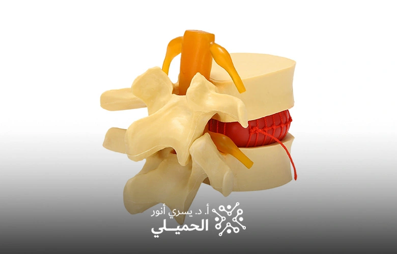
Lumbar Disc Removal Using a Surgical Microscope
The discs in the lumbar region of the spine can be precisely and effectively treated using this technique, which minimizes risks and reduces recovery time.
Lumbar disc removal using a surgical microscope is a procedure where the herniated or damaged part of the lumbar disc is removed with the assistance of a surgical microscope. This microscope provides high magnification and a wide field of view for the surgeon, facilitating precise work and minimizing interference with surrounding healthy tissues.
A surgical microscope assists the spine surgeon in visualizing the affected disc and surrounding tissues, allowing precise removal of the herniated disc portion under the nerve root. By creating more space for the nerve root, pressure is relieved. Since microdiscectomy is performed through small incisions, pain and complications are minimized, leading to much faster recovery and a quicker return to daily activities. Additionally, microdiscectomy offers a lower chance of disc herniation recurrence. The procedure steps include:
If you are suffering from pain or issues in the lumbar region of your back, contact Dr. Yousry El-Hamili today to book your consultation with our specialized medical team and discover how we can help you receive the best treatment.